Imaging of Signaling Events
Participants: Fahrbach, Guthold, Johnson, King, Macosko, McCauley, Muday, Pratt, Poole, Silver
Questions: This group has coalesced around a common need for microscopic imaging tools to study molecular communication events including recognition of environmental cues, response by activating intracellular mechanisms, and communication of this information to nearby and/or distant cells.

Areas of research focus include hormonal signaling in plants, insects and vertebrates, G-coupled protein receptors, stress-physiology pathways, oxidation and post-translational modification, insulin-related signaling and secretion, mechanisms of transport within cells, and the diffusion and binding of DNA repair proteins.
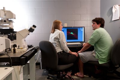
Technology: Visualization of molecular localization and signaling with high resolution in living cells is now possible. Laser scanning confocal microscopy (LSCM), which allows imaging of thin optical sections from thick samples, has been critical to this advancement. LSCM coupled with fluorescent labels (in particular the Nobel Prize winning green fluorescent protein) permits the visualization of multiple receptors, intracellular signaling proteins, and transcription factors in living cell populations. Center participants, led by McCauley, have collaborated successfully on three imaging equipment proposals in the past five years. One award, enabling purchase of a LSCM, has revolutionized signaling research at WFU; research productivity has increased, resulting in seven publications, and WFU has been established as a leader in imaging, evidenced by hosting distinguished speakers, regional workshops, and visiting scientists. Greater technology is still needed to advance the ambitious goals of the center participants. A multi-departmental group of researchers, all center participants, have submitted an NSF application to meet some urgent imaging needs (McCauley, Johnson, Muday, Bonin, and King, PI and CoPIs; $258,251 requested to increase capabilities on the LSCM). Yet other needs remain, including integration of a drug-delivery system on the LSCM and a dedicated live-cell imaging confocal system. Participants in this group are building on the legacy of successful equipment collaborations to develop greater intellectual collaborations which bridge disciplinary boundaries. For example, McCauley and Johnson recently jointly developed a method for analyzing changes in protein localization (50).
Emphasis Group Activities: Critical to successful imaging research is expert data acquisition and analysis. Members of this group have significant expertise in diverse areas of imaging, including calcium imaging, high-throughput screening, photomanipulation and particle tracking. Monthly meetings, organized by McCauley, will focus on working issues, including techniques, ongoing projects, and future experiments, fostering development of collaborations based upon common interests and complementary skills. Activities of this subgroup are organized by Anita McCauley.
Implications: The collaborative projects within this group investigate how chemotherapeutic agents kill cells, how proteins find and repair potentially cancer-causing mutations, and how pain or environmental stress is signaled in organisms. Center support will facilitate studies of how hormones and the environment affect the growth and development of agriculturally important crop species, efforts to decipher the cellular basis for learning and memory, greater use of high throughput, high content imaging, and further establish WFU as a leader in microscopic imaging.
